Dot and Blot Hemorrhage
![Dot and Blot Hemorrhages](https://www.vagelos.columbia.edu/sites/default/files/styles/cola_media_200/public/media/images/2022-03/dot-and-blot-hemorrhage.jpeg?itok=mACVTOQG 200w, https://www.vagelos.columbia.edu/sites/default/files/styles/cola_media_260/public/media/images/2022-03/dot-and-blot-hemorrhage.jpeg?itok=e2p8XiD6 260w, https://www.vagelos.columbia.edu/sites/default/files/styles/cola_media_320/public/media/images/2022-03/dot-and-blot-hemorrhage.jpeg?itok=b9t5BhKx 320w, https://www.vagelos.columbia.edu/sites/default/files/styles/cola_media_400/public/media/images/2022-03/dot-and-blot-hemorrhage.jpeg?itok=U2BHCo_g 400w, https://www.vagelos.columbia.edu/sites/default/files/styles/cola_media_1600/public/media/images/2022-03/dot-and-blot-hemorrhage.jpeg?itok=9donPYR4 430w, https://www.vagelos.columbia.edu/sites/default/files/styles/cola_media_520/public/media/images/2022-03/dot-and-blot-hemorrhage.jpeg?itok=nI22L07d 520w, https://www.vagelos.columbia.edu/sites/default/files/styles/cola_media_640/public/media/images/2022-03/dot-and-blot-hemorrhage.jpeg?itok=pSh0x-8R 640w, https://www.vagelos.columbia.edu/sites/default/files/styles/cola_media_800/public/media/images/2022-03/dot-and-blot-hemorrhage.jpeg?itok=UEWAtbbI 800w, https://www.vagelos.columbia.edu/sites/default/files/styles/cola_media_1040/public/media/images/2022-03/dot-and-blot-hemorrhage.jpeg?itok=J8jZSe9U 1040w, https://www.vagelos.columbia.edu/sites/default/files/styles/cola_media_1280/public/media/images/2022-03/dot-and-blot-hemorrhage.jpeg?itok=QgCkjIdB 1280w)
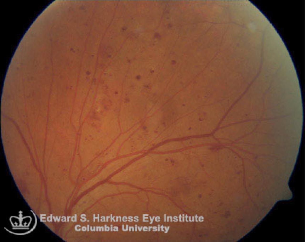
- Hemorrhages lie deeper in the retina.
- Usually blood accumulates in the outer plexiform or inner nuclear layers, or more easily seen at peripheral retina where the nerve fiber layer is thin.
- Commonly seen in association with diabetic or hypertensive retinopathy, peripapillary hemorrhage in patients with normal tension glaucoma that is also called splinter hemorrhage and retinal vein occlusion.