You are here
- Home >
- Departments & Centers >
- Ophthalmology >
- Research >
- Research Labs >
- John Lab >
- Lab Members
Lab Members
Research Scientists
Christa Montgomery, PhD
- Research Scientist
I have been the Lab Manager and a Research Scientist in the Simon John Lab since January of 2019. I earned my interdisciplinary Ph.D. in Molecular Biology and Biochemistry with a co-discipline Cell Biology and Biophysics from the University of Missouri at Kansas City in 2011. I have experience with protein biochemistry and metabolism, transformed and primary cell culture and assays, and have studied mouse and rat models of autoimmune optic neuritis, metabolic syndrome/diabetes, hyperlipidemia, and glaucoma.

Chi Zhang, PhD
- Research Scientist
I have been an associate research scientist in John Lab since February of 2019. I earned my Ph.D. in Developmental Biology from Fudan University in 2012 where I used different mouse models to investigate heart, kidney and retinal diseases. Since joining the John lab, my focus has been on developing inducible glaucoma mouse models as well as molecular mechanism of neural damage in glaucoma.

Research Assistants, Associates, Aides & Graduate Students
Aakriti Bhandari, BS
- Research Assistant
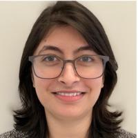
I joined the John Lab in 2018 as a research intern after receiving my B.S in Biochemistry and Neuroscience from Centenary College of Louisiana. I have experience with animal husbandry and genotyping, physiology, immunohistochemistry, and microscopy.

Logan Horbal, BS
- Research Assistant
I joined the John lab in September of 2018 as a research assistant. I earned my BS in Physiology & Neurobiology from the University of Connecticut in 2017. I have experience with behavioral assays, ocular phenotyping of mice, immunohistochemistry, data visualization with R, and computer-aided design.

Sally Zhou, BS
- Research Associate I
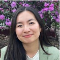
I joined the John Lab as a research assistant after I graduated from Northeastern University in May of 2021 with a B.S. in Biology. In my previous research experience, I studied the mutational frequency of mice treated with NDMA, a water contaminant that is known to cause DNA adducts that contribute to the development of cancer in mouse models. During the pandemic, I worked at Massachusetts General Brigham’s COVID-19 Diagnostic Accelerator Lab to validate alternative COVID-19 diagnostic devices. At the John Lab, I am excited to be exposed to working with animals models, studying mechanisms of ocular disease, and various molecular and physiological techniques such as IHC, ISH, dissection, and genotyping. I believe this experience will enhance my foundation in science and support my journey to pursue a career in medicine. In my free time, I enjoy cooking meals from various cultures and I plan to adopt a cat soon.

Felicia Juarez, BS
- Research Associate I
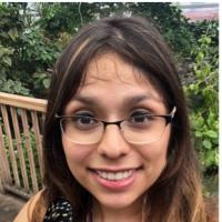
I received my Bachelor of Science in Neuroscience with a concentration in Molecular and Cellular Biology from the Johns Hopkins University in 2017. My main research interests are genetics and neuroscience. As an undergraduate, I worked in Seth Blackshaw's lab on a project entitled "Investigating the role of Lhx2 in the development of the ciliary body." Since graduating college, I have worked in several labs ranging from studying blood diseases to building a library of nuclear constructs to study the phase separation of nuclear proteins in vivo. I am experienced in tissue culture, mouse husbandry, cloning, and various molecular biology techniques. I joined the John lab in June of 2020 as a Research Associate. During my free time I enjoy reading, exploring new areas in New York, and spending time with my dog, Nova.

Tracy Preko, BS
- Research Aide
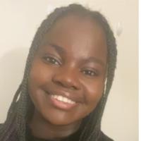
I joined the John Lab as a research aide after I graduated from the University of Notre Dame in May of 2021 with a B.S. in Neuroscience and Behavior along with a minor in Poverty Studies. During my time here I am excited to gain in-depth knowledge into the development of glaucoma and how it is investigated in mice models while gaining experience in various techniques and procedures like genotyping, animal husbandry, and immunohistochemistry. My hobbies include exploring new recipes and dishes while catching up on my favorite shows and movies.

Nicholas Tolman, BA
- Graduate Student
I received my B.A. in Behavioral Neuroscience from Connecticut College . During my time at Connecticut College, I worked on several different research projects. We studied whether Ceftriaxone and other β-lactam antibiotic compounds could attenuate morphine reward in rats by enhancing the reuptake rate of excitatory amino-acid transporters. For my honors thesis, I examined the effects of developmental lead exposure on working memory performance in rats after manipulating several elements of the animal’s developmental environment.
I started my Ph.D. in the John lab in 2016. I am studying the roles of a LIM domain transcription factor , LMX1b, in ocular development and adult-onset glaucoma. This has allowed me an unusually broad graduate training including clinical, molecular, genetic, genomic, and other computational/statistical approaches using R, Seurat and other software. Beyond immunohistochemistry and other standard molecular techniques, I have mastered various delicate dissections (retina, optic nerve, brain regions and ocular drainage tissues) and precise ocular physiological measurements (including intraocular pressure and ocular fluid drainage or outflow). My projects have also involved clinical and histologic examination of mouse eyes and Mendelian and complex genetics including the diversity outbred mouse population. I have been involved with several other exciting projects, including the screening for and characterization of high IOP mutants and in a collaboration with engineers towards developing new treatment tools. Currently, I am focusing on the developmental biology of ocular development in Lmx1b mutants as well as the gene expression networks controlling ocular development and IOP elevation (using single cell sequencing, bulk RNA sequencing and other approaches). Dr. John has facilitated my interaction with collaborators and exposed me to grant writing, lab. management decisions and strategic planning. This diverse training along with his mentorship and the lab's environment are putting me in a strong position for my future career as a PI.
Being from coastal Maine, I was used to being an outdoor person at our lab's initial location next to Acadia National Park. The exciting and fast paced environment at our new location, Columbia University, is an amazing experience with the opportunity to learn from many first-rate scientists.

Jocelyn Thomas, BA
- Research Assistant
Science, and more specifically, life science, has been a passion of mine since an early age. I continued to foster that curiosity as an undergraduate at Colby College where I received my B.A. in Biology. I was also part of the Colby Achievement Program in the Sciences. While at Colby, I had the opportunity to participate in research in several laboratories, with projects ranging from measuring the effects of an acetylcholine rich diet on the development of the hippocampus portion of rat brains, to better understanding the mechanisms how circadian rhythm proteins cause their effects in fiddler crabs. I joined the John Laboratory team a few months after graduating and I have already learned so much during my time here. I have learned a vast array of techniques for both handling and collecting data from the mice we work with. I have also been trained to carryout genotyping, as well as optic nerve dissections. I really enjoy working in the John Laboratory and appreciate the chance to continue to develop my scientific research skills. The latter is something the John lab is invested in doing for their RAs.
When I am not in the lab I am out getting to know Bar Harbor better. Even though I am a native Mainer, I have only been to Bar Harbor a handful of times, but I truly enjoy the outdoors and I look forward to exploring more of this beautiful area.

Kelly Keezer
- Laboratory Technician IV- next: The Howell Lab, The Jackson Laboratory, Bar Harbor, ME
I have been a Laboratory Technician IV in the lab since 2014. I help maintain our core mouse colonies that are needed by everyone in the lab to complete experiments. I am also involved in research with one of our postdoctoral fellows, to develop an inducible model of glaucoma in mice. Our goal is to raise intraocular pressure and cause optic nerve degeneration, as seen in glaucoma. I perform intraocular injections and follow up by measuring intraocular pressure (IOP) using a tonometer and evaluate optic nerve cupping using optical coherence tomography (OCT). An inducible model of glaucoma will allow us to more quickly assess whether mutations and treatments affect the course of the disease.
I really enjoy working in the lab, as the group continually challenges me to master new techniques. Despite having two very active kids, I am currently working towards my Bachelors Degree through Husson University during the evenings.
![Kelly Keezer]()
Administrative Staff
Lisa Marie Izquierdo, MA
- Administrative Coordinator
I joined the John Lab in October 2019 as the team's Administrative Coordinator. I received my Master’s in International Affairs from The New School in New York City, NY and previously spent 8 years at New York City Health + Hospitals/Bellevue. Since joining the lab, I have enjoyed learning about research administration and the many advances the John Lab is making in addressing the causes of glaucoma.

Bethany Preble, BS
- Executive Administrative Assistant
In 2007, joined the John Lab as Simon's Executive Administrative Assistant with the Howard Hughes Medical Institute. Prior to the lab, I worked in management at a local YMCA, as a training coordinator for a state agency, and in numerous off-shoots of both. I am originally from Maine and have two wonderful children. Science has taken on a new role in my life, and watching the lab make great things happen has been a super opportunity.

Visiting Investigators
Jeffrey Marchant, PhD
- Research Assistant Professor, Department of Anatomy and Cellular Biology, Tufts University
Dr. Marchant is a Research Assistant Professor at Tufts University School of Medicine and has been studying the vertebrate eye since graduate school. Much of the early work focused on the cornea and currently he is funded to develop a cornea replacement using silk scaffolds that have been seeded with human corneal cells. His research expanded to begin a characterization of the corneoscleral angle in the chicken and to study trabecular meshwork cells using human cell cultures. In 2005, Dr. Marchant began collaborating with Dr. John to further these studies by switching to mouse models of IOP regulation. The research has led to the discovery of a novel basement membrane specialization that appears to support Schlemm’s canal giant vacuoles throughout their expansion cycle. This specialization may also serve as a dynamic modulator of aqueous outflow resistance. Through the collaboration with Dr. John,
Dr. Marchant has obtained external funding from the American Health Assistance Foundation - National Glaucoma Research. Dr. Marchant continues to spend time in Dr. John’s laboratory as a Visiting Investigator each summer. At Tufts, he can be reached at jeffrey.marchant@tufts.edu(link sends e-mail).

James Morgan, PhD
- Professor, School of Optometry & Vision Sciences, Cardiff University
Dr. Morgan's research is aimed at understanding the structural and cellular changes occurring at the level of the optic nerve in glaucoma using clinical and laboratory-based techniques. Glaucoma is one of the most common causes of vision loss in the UK population, affecting up to 2% of the population. Classically, it is associated with increased intraocular pressure, which causes the death of retinal ganglion cells and results in a slow and, if untreated, progressive loss of vision. In most cases, peripheral vision is lost first and is usually asymptomatic. By the time a patient notices restriction of visual field, advanced damage may have occurred to the optic nerve that is usually irreversible. Treatment is aimed at the reduction of eye pressure (intraocular pressure) to prevent this vision loss. Usually, this is achieved using eye drops although, in some cases, surgery or even laser treatment may be required. One of the key problems is in identifying the disease in its earliest stages and in detecting the first signs of retinal (optic nerve) damage. As a consultant ophthalmologist, Morgan runs a glaucoma service at the University Hospital of Wales. He currently chairs the Expert Working Group for the development of shared care glaucoma in Wales where they are working to develop the role of virtual clinics for the community care of patients with diagnosed glaucoma. His email address is MorganJE3@cardiff.ac.uk(link sends e-mail).
Clinical Studies:
These are based at the Retinal Imaging Laboratory and are directed at the quantification of structural optic nerve changes in the early stages of glaucoma. Since these precede the development of visual field changes as detected using clinical perimetric techniques, their detection should enable early disease diagnosis and improve the long-term prognosis for the patient. Morgan's lab is currently assessing the value of digital stereoviewing in the quantification of these nerve head changes. In addition, his lab is assessing the value of other imaging modalities such as OCT in the determination of optic nerve head changes that occur in early glaucoma.
Using a variety of in vivo and in vitro models of glaucoma, Morgan's group is investigating the mechanisms of retinal ganglion cell death in glaucoma. There is good evidence that these cells undergo a prolonged period of atrophy and remodeling prior to cell death, which is manifest as shrinkage and pruning in the cell body and dendritic processes. These changes will reduce the efficiency with which these cells can detect visual signals but also suggests that this damage could be reversed. They use techniques such as biolistics transfection to see if the overexpression of genes that might be beneficial for retinal neurons can result in cell rescue Retinal ganglion cell transfected with plasmid coding for GFP. Retinal Explant preparation after 24 hours in culture.

Pedro P. Irazoqui, PhD
- Associate Head of Biomedical Engineering, Showalter Faculty Scholar, Associate Professor of Electrical and Computer Engineering, Director, Center for Implantable Devices, Purdue Universit
Dr. Irazoqui received his BSc and MSc degrees in Electrical Engineering from the University of New Hampshire, Durham in 1997 and 1999 respectively, and the Ph.D. in Neuroengineering from the University of California at Los Angeles in 2003.Currently he is an associate professor in the Weldon School of Biomedical Engineering at Purdue University, and director of the Center for Implantable Devices pursuing research into a modular approach to the design of biological implants. Devices are applied to the clinical treatment of physiological disorders, using miniature, wireless, implantable systems. Specific research and clinical applications explored include: epilepsy, glaucoma, cardiology, and neural interfaces.He has received the Best Teacher Award from the Weldon School of Biomedical Engineering (2006 & 2009), the Early Career Award from the Wallace H. Coulter Foundation (2007 & Phase II in 2009), the Marion B. Scott Excellence in Teaching Award from Tau Beta Pi (2008), and the Outstanding Faculty Member Award from the Weldon School of Biomedical Engineering (2009), and has been serving as Associate Editor of IEEE Transactions on Biomedical Engineering since late 2006.
Research Interests
- Application, design, and fabrication of implantable analog integrated circuits
- High speed RF circuits for wireless data and power coupling to and from biological implants
- Real-time discrete-time signal processing for biological signals
- Neuronal substrates of behavior, perception, and locomotion
![Pedro P. Irazoqui]()


